By Truth | The Healers Journal
—
2013 was an incredible year for neuroscience in which a number of truly groundbreaking studies revealed many new insights about the inner workings of our minds. Some of these discoveries in and of themselves are significant enough to create entirely new fields of research within the larger field of neuroscience.
What’s truly remarkable about the studies listed below is that their findings are quite tangible for anyone looking to ‘optimize’ their lives and better understand how their own minds work. Simply by browsing through the summaries of the research it is possible to find many ways you can immediately begin to substantially improve your life. Perhaps this is why neuroscience is such an exciting field — the resulting data is typically extremely practical and leads to truly actionable items we can immediately use to create better lives for ourselves.
1. Human Brains Are Hardwired for Empathy, Friendship
Perhaps one of the most defining features of humanity is our capacity for empathy — the ability to put ourselves in others’ shoes. A new University of Virginia study strongly suggests that we are hardwired to empathize because we closely associate people who are close to us — friends, spouses, lovers — with our very selves.”With familiarity, other people become part of ourselves,” said James Coan, a psychology professor in U.Va.’s College of Arts & Sciences who used functional magnetic resonance imaging brain scans to find that people closely correlate people to whom they are attached to themselves. The study appears in the August issue of the journal Social Cognitive and Affective Neuroscience.
“Our self comes to include the people we feel close to,” Coan said.
In other words, our self-identity is largely based on whom we know and empathize with. Coan and his U.Va. colleagues conducted the study with 22 young adult participants who underwent fMRI scans of their brains during experiments to monitor brain activity while under threat of receiving mild electrical shocks to themselves or to a friend or stranger. The researchers found, as they expected, that regions of the brain responsible for threat response — the anterior insula, putamen and supramarginal gyrus — became active under threat of shock to the self. In the case of threat of shock to a stranger, the brain in those regions displayed little activity. However when the threat of shock was to a friend, the brain activity of the participant became essentially identical to the activity displayed under threat to the self.
“The correlation between self and friend was remarkably similar,” Coan said. “The finding shows the brain’s remarkable capacity to model self to others; that people close to us become a part of ourselves, and that is not just metaphor or poetry, it’s very real. Literally we are under threat when a friend is under threat. But not so when a stranger is under threat.”
Coan said this likely is because humans need to have friends and allies who they can side with and see as being the same as themselves. And as people spend more time together, they become more similar.
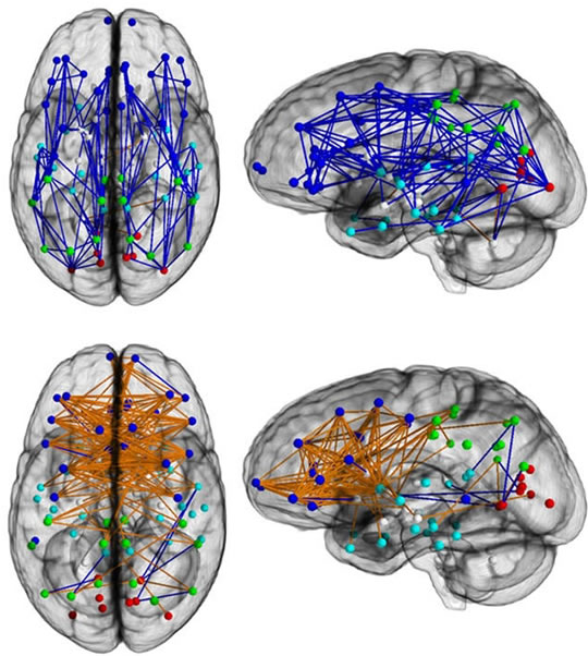
2. Brain Connectivity Study Reveals Striking Differences Between How Men and Women’s Brains are Wired

Male brains (top) show greater connectivity front-to-back, while female brains (bottom) are more connected across the hemispheres. Source: Science Daily
A new brain connectivity study from Penn Medicine published today in the Proceedings of National Academy of Sciences found striking differences in the neural wiring of men and women that’s lending credence to some commonly-held beliefs about their behavior.
In one of the largest studies looking at the “connectomes” of the sexes, Ragini Verma, PhD, an associate professor in the department of Radiology at the Perelman School of Medicine at the University of Pennsylvania, and colleagues found greater neural connectivity from front to back and within one hemisphere in males, suggesting their brains are structured to facilitate connectivity between perception and coordinated action. In contrast, in females, the wiring goes between the left and right hemispheres, suggesting that they facilitate communication between the analytical and intuition.
“These maps show us a stark difference–and complementarity–in the architecture of the human brain that helps provide a potential neural basis as to why men excel at certain tasks, and women at others,” said Verma.
For instance, on average, men are more likely better at learning and performing a single task at hand, like cycling or navigating directions, whereas women have superior memory and social cognition skills, making them more equipped for multitasking and creating solutions that work for a group. They have a mentalistic approach, so to speak.
3. Hidden Caves in the Brain Open Up During Sleep to Wash Away Toxins
Sleep hoses garbage out of the brain, a study of mice finds.
The trash, including pieces of proteins that cause Alzheimer’s disease, piles up while the rodents are awake. Sleep opens spigots that bathe the brain in fluids and wash away the potentially toxic buildup, researchers report in the Oct. 18 Science.
The discovery may finally reveal why sleep seems mandatory for every animal. It may also shed new light on the causes of neurodegenerative disorders such as Alzheimer’s and Parkinson’s diseases.
“It’s really an eye-opening and intriguing finding,” says Chiara Cirelli, a sleep researcher at the University of Wisconsin–Madison. The results have already led her and other sleep scientists to rethink some of their own findings.
Although sleep requirements vary from individual to individual and across species, a complete lack of it is deadly. But no one knows why.
To test the detox power of sleep, the group demonstrated that a protein fragment called β-amyloid, located in the interstitial space, disappeared faster while animals were sleeping. β-amyloid is best known as a component of amyloid plaques in association with Alzheimer’s disease.
Commenting on the research, the lead scientist, Maiken Nedergaard, the co-director of the Center for Translational Neuromedicine at the University of Rochester Medical Center in New York, said in a research note: “Sleep changes the cellular structure of the brain. It appears to be a completely different state.”
4. Researchers Debunk Myth of ‘Right-Brained’ and ‘Left-Brained’ Personality Traits
Chances are, you’ve heard the label of being a “right-brained” or “left-brained” thinker. Logical, detail-oriented and analytical? That’s left-brained behavior. Creative, thoughtful and subjective? Your brain’s right side functions stronger — or so long-held assumptions suggest.
But newly released research findings from University of Utah neuroscientists assert that there is no evidence within brain imaging that indicates some people are right-brained or left-brained.
For years in popular culture, the terms left-brained and right-brained have come to refer to personality types, with an assumption that some people use the right side of their brain more, while some use the left side more.
Following a two-year study, University of Utah researchers have debunked that myth through identifying specific networks in the left and right brain that process lateralized functions. Lateralization of brain function means that there are certain mental processes that are mainly specialized to one of the brain’s left or right hemispheres. During the course of the study, researchers analyzed resting brain scans of 1,011 people between the ages of seven and 29. In each person, they studied functional lateralization of the brain measured for thousands of brain regions — finding no relationship that individuals preferentially use their left -brain network or right- brain network more often.
“It’s absolutely true that some brain functions occur in one or the other side of the brain. Language tends to be on the left, attention more on the right. But people don’t tend to have a stronger left- or right-sided brain network. It seems to be determined more connection by connection, ” said Jeff Anderson, M.D., Ph.D., lead author of the study, which is formally titled “An Evaluation of the Left-Brain vs. Right-Brain Hypothesis with Resting State Functional Connectivity Magnetic Resonance Imaging.” It is published in the journal PLOS ONE this month.
5. Sleeping Patterns Shape Our Brains: Different Neural Structures Found in the Brains of Night Owls
A recent study by researchers in Germany has suggested that night owls have a decreased amount of white matter in their brains compared to people who go to bed early. White matter is insulated and transmits nerve signals around the brain. In this study three classes of chronotypes were analysed; early (EC), intermediate (IC) and late (LC). Chronotype simply refers to the body clock of an individual, the time physical functions or change occur (eg; eating, body temperature or this case sleeping).
ECs wake up early and go to bed early where as LCs go to bed late and sleep into the day. There are many factors that are believed to contribute to the sleep chronotype of an individual. Young adults with high testosterone levels (mainly males) are more likely to go to bed late where as those with low testosterone levels (mainly females) are more likely to go to be earlier. This preference also varies with age, young people tend to sleep in where as elderly people wake up earlier.
Why is this important?
Well it has been shown that depressed individuals exhibit abnormal white matter levels in their brains, specifically in the anterior cingulate gyrus (ACC) and the corpus callosum. The study showed a correlation between chronotype and white matter composition in the left ACC corpus callosum.
LCs were found to have decreased amounts of white matter in these regions compared to ECs and ICs. It is thought that the decreased white matter due to their lifestyle choices puts them at a higher risk of depression amongst other bipolar disorders.
6. Direct Brain to Brain Communication in Humans
We sought to demonstrate that it is possible to send information extracted from one brain directly to another brain, allowing the first subject to cause a desired response in the second subject through direct brain-to-brain communication. A task was designed such that the two subjects could cooperatively solve the task by transmitting a meaningful signal from one brain to the other.
The experiment leverages two existing technologies: electroencephalography (or EEG) for noninvasively recording brain signals from the scalp and transcranial magnetic stimulation (or TMS) for noninvasively stimulating the brain.
The task that the subjects must cooperatively solve via brain-to-brain communication is a computer game (Figure 4). The task involves saving a “city” (on the left) from getting hit by rockets fired by a “pirate ship” from the lower right portion of the screen (depicted by skull-and-bones). To save the city, the subjects must fire a “cannon” located at the lower center portion of the screen. If the “fire” button is pressed before the moving rocket reaches the city, the rocket is destroyed (Figure 5), the city is saved, and the trial ends. To make the task more interesting, on some trials, a friendly “supply plane” may appear instead of a pirate rocket and move leftwards towards the city (Figure 6). The subjects must avoid firing the cannon at the supply plane.
The results suggest that information extracted noninvasively from one brain using EEG can be transmitted to another brain noninvasively using TMS to allow two persons to cooperatively solve a task via direct brain-to-brain transfer of information. The next phase of the study will attempt to quantify this transfer of information using a larger pool of human subjects.
7. Sound Waves Proven to be Able to Affect Human Mood
University of Arizona researchers have found in a recent study that ultrasound waves applied to specific areas of the brain appear able to alter patients’ moods.
The technique could one day be used to treat conditions such as depression and anxiety.
With research committee and hospital approval, and patient informed consent, Dr. Stuart Hameroff, professor emeritus of the UA’s departments of anesthesiology and psychology and director of the UA’s Center for Consciousness Studies, and colleagues applied transcranial ultrasound stimulation (TUS) to 31 chronic pain patients at The University of Arizona Medical Center-South Campus.
This was a double-blind study (neither doctor nor subject knew if the ultrasound machine had been switched on or off).
Sites for transcranial ultrasound (TUS). 1-4 are used in Doppler blood fl ow studies. 1: trans-orbital, 2: sub-mandibular, 3: sub-occipital, 4: temporal window. 5 is the site used in present study, overlying frontal-temporal cortex. (Credit: Stuart Hameroff et al./Brain Stimulation)
Patients reported improvements in mood for up to 40 minutes following treatment with brain ultrasound, compared with no difference in mood when the machine was switched off.
The researchers confirmed the patients’ subjective reports of increases in positive mood with a Visual Analog Mood Scale, or VAMS, a standardized objective mood scale often used in psychological studies.
8. Brain Releases Natural Painkillers During Social Rejection
“Sticks and stones may break my bones, but words will never hurt me,” goes the playground rhyme that’s supposed to help children endure taunts from classmates. But a new study suggests that there’s more going on inside our brains when someone snubs us — and that the brain may have its own way of easing social pain.
The findings, recently published in Molecular Psychiatry by a University of Michigan Medical School team, show that the brain’s natural painkiller system responds to social rejection — not just physical injury.
What’s more, people who score high on a personality trait called resilience — the ability to adjust to environmental change — had the highest amount of natural painkiller activation.
The team, based at U-M’s Molecular and Behavioral Neuroscience Institute, used an innovative approach to make its findings. They combined advanced brain scanning that can track chemical release in the brain with a model of social rejection based on online dating. The work was funded by the U-M Depression Center, the Michigan Institute for Clinical and Health Research, the Brain & Behavior Research Foundation, the Phil F Jenkins Foundation, and the National Institutes of Health.
They focused on the mu-opioid receptor system in the brain — the same system that the team has studied for years in relation to response to physical pain. Over more than a decade, U-M work has shown that when a person feels physical pain, their brains release chemicals called opioids into the space between neurons, dampening pain signals.
9. Your Brain ‘Sees’ Things Even When You Don’t
University of Arizona doctoral degree candidate Jay Sanguinetti has authored a new study, published online in the journalPsychological Science, that indicates that the brain processes and understands visusal input that we may never consciously perceive.
The finding challenges currently accepted models about how the brain processes visual information.
A doctoral candidate in the UA’s Department of Psychology in the College of Science, Sanguinetti showed study participants a series of black silhouettes, some of which contained meaningful, real-world objects hidden in the white spaces on the outsides.
Saguinetti worked with his adviser Mary Peterson, a professor of psychology and director of the UA’s Cognitive Science Program, and with John Allen, a UA Distinguished Professor of psychology, cognitive science and neuroscience, to monitor subjects’ brainwaves with an electroencephalogram, or EEG, while they viewed the objects.
“We were asking the question of whether the brain was processing the meaning of the objects that are on the outside of these silhouettes,” Sanguinetti said. “The specific question was, ‘Does the brain process those hidden shapes to the level of meaning, even when the subject doesn’t consciously see them?”
The answer, Sanguinetti’s data indicates, is yes.
Study participants’ brainwaves indicated that even if a person never consciously recognized the shapes on the outside of the image, their brains still processed those shapes to the level of understanding their meaning.


